Energy levels in atoms
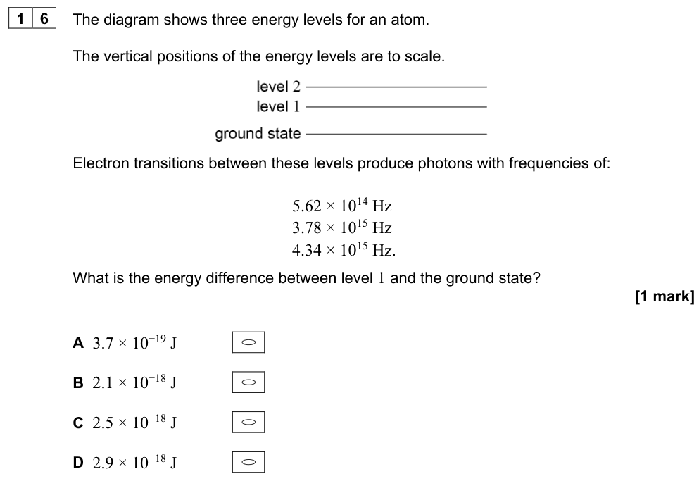
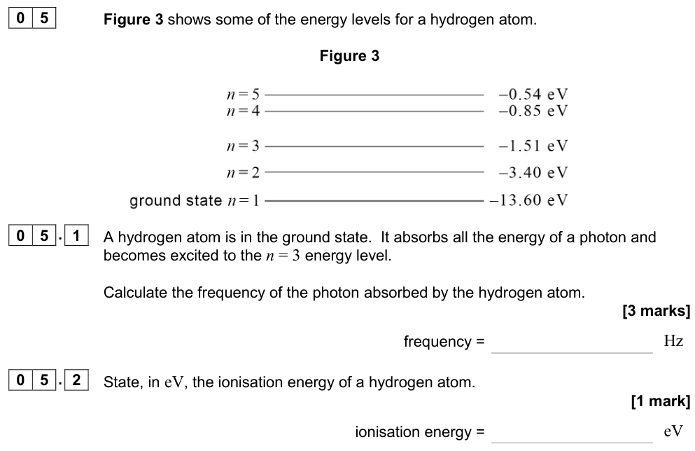
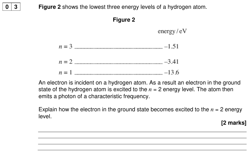
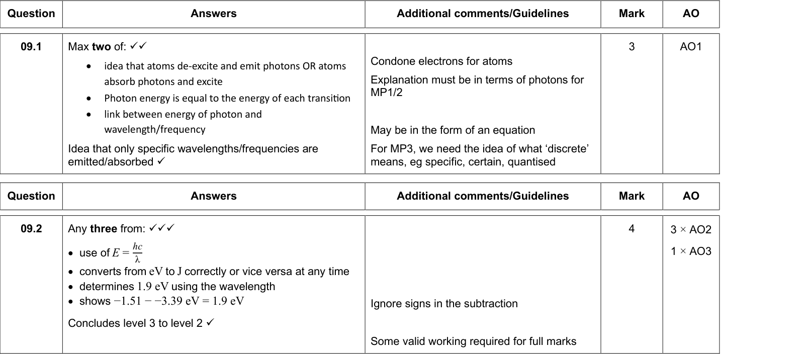
The electrons in an atom can only exist in certain discrete energy levels. They cannot have energies in between.
- The lowest level is the ground state. The electron is most stable there.
- Higher levels are excited states.
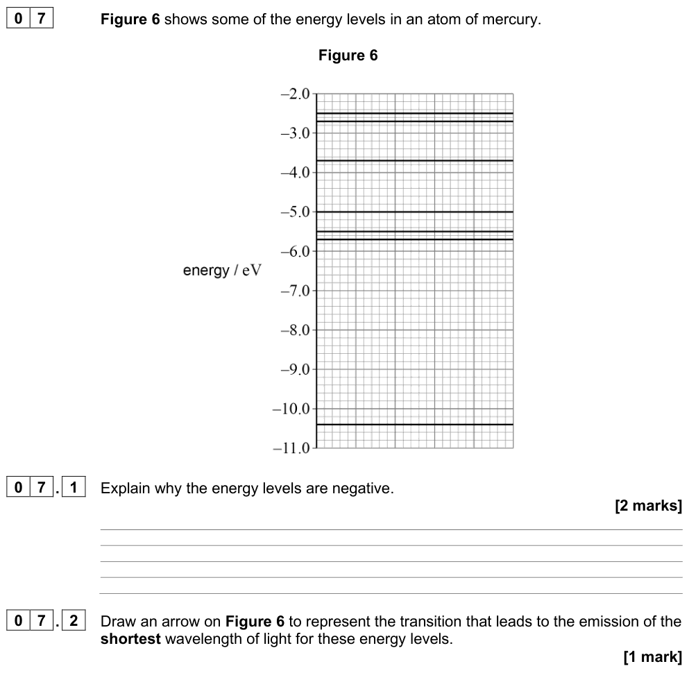
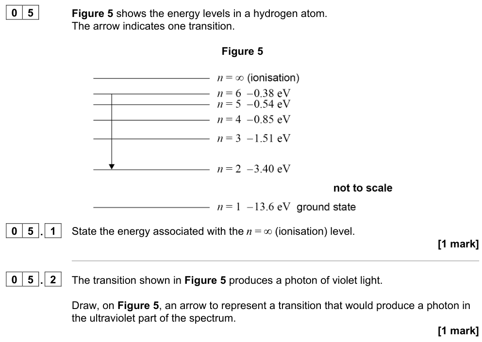
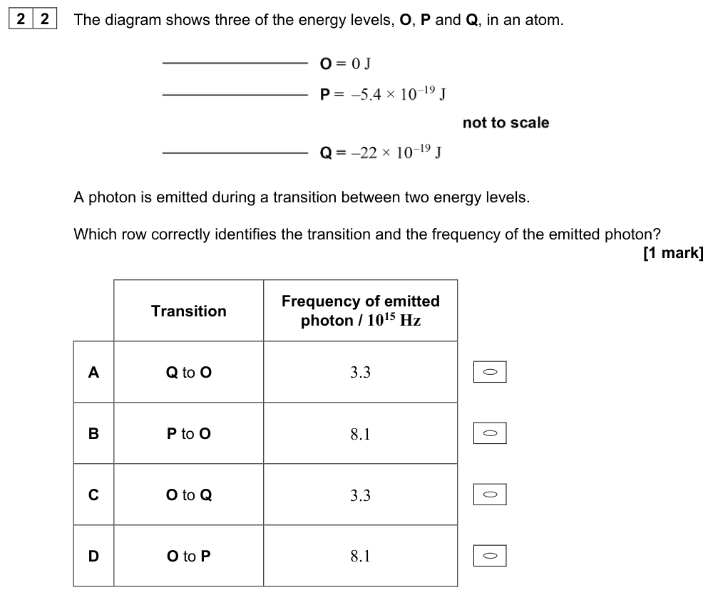
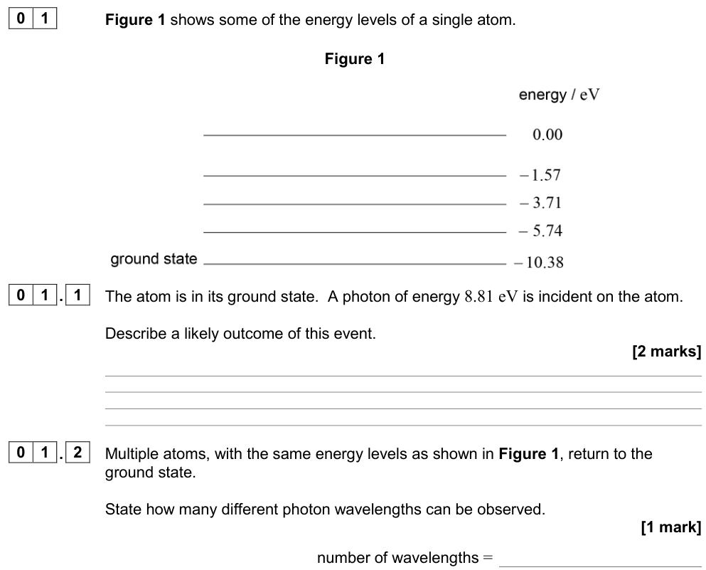
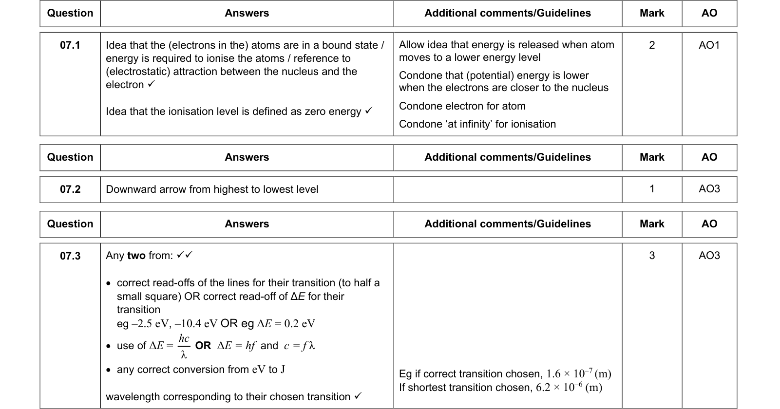
- Energy levels are usually given as negative values in eV. The energy of an electron that has just escaped from the atom (at rest, at infinity) is defined as zero. An electron in the atom is bound, so it has less energy than this: negative energy. The ionisation level is $E = 0$.
Excitation and ionisation
Excitation and ionisation
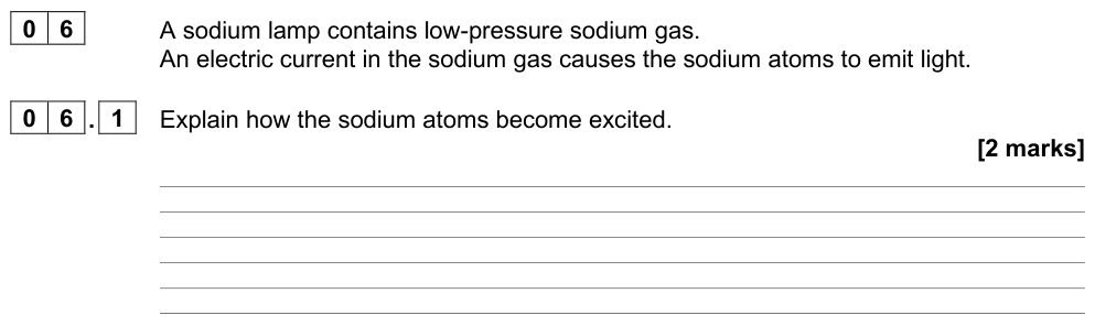
Excitation: an electron in an atom absorbs energy and moves up to a higher energy level (but stays in the atom).
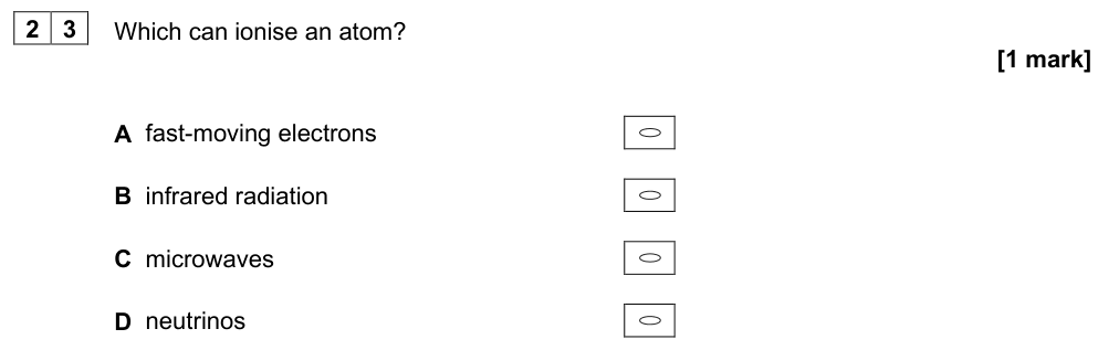
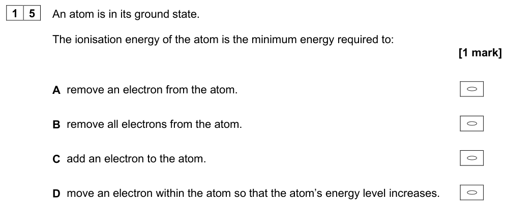
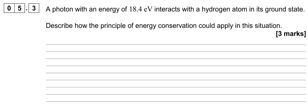
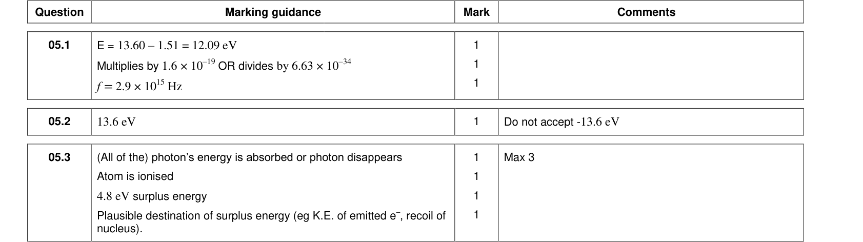
Ionisation: an electron absorbs enough energy to escape completely from the atom, leaving a positive ion. The ionisation energy is the minimum energy needed to remove an electron from the ground state (13.6 eV for hydrogen).
Atoms can be excited in two ways:
- By photons: a photon is absorbed only if its energy exactly equals the difference between two energy levels (unless it has enough energy to ionise the atom). The whole photon is absorbed or none of it.
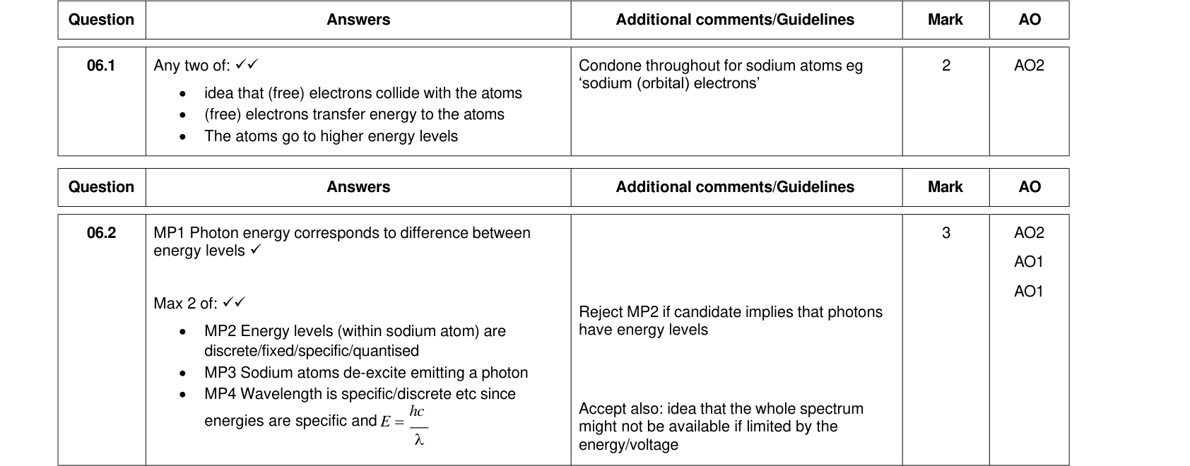
- By colliding electrons: a free electron can give part of its kinetic energy to an atomic electron. It needs at least enough energy to make the transition; any excess stays with the colliding electron as kinetic energy.
De-excitation and photon emission
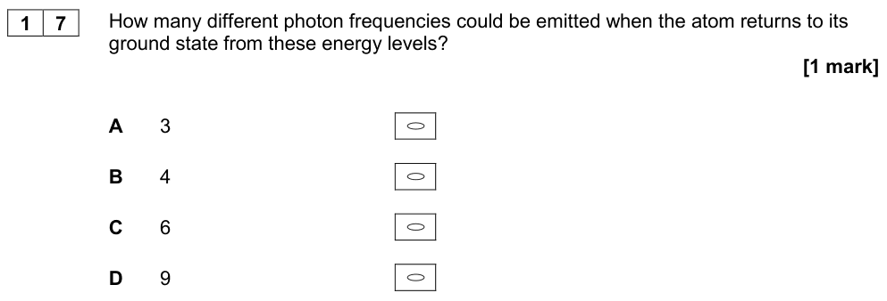
Excited states are unstable. The electron drops back down to a lower level, in one or several steps, and emits a photon for each step:
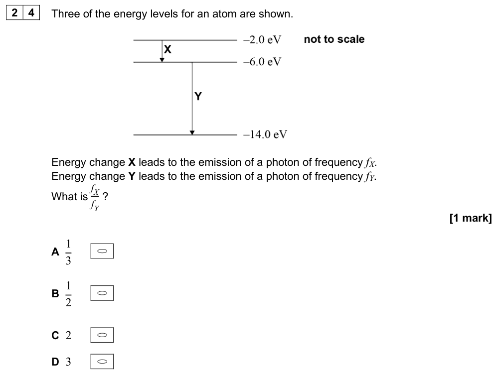
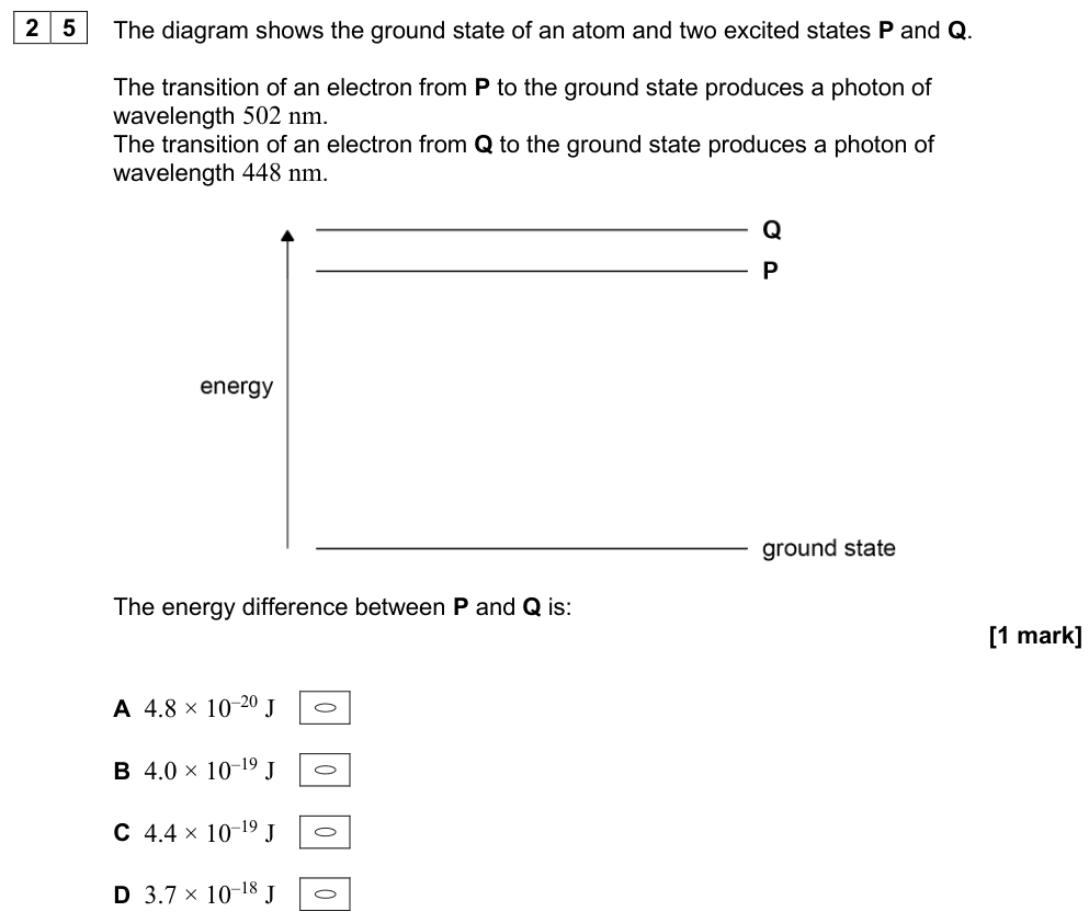
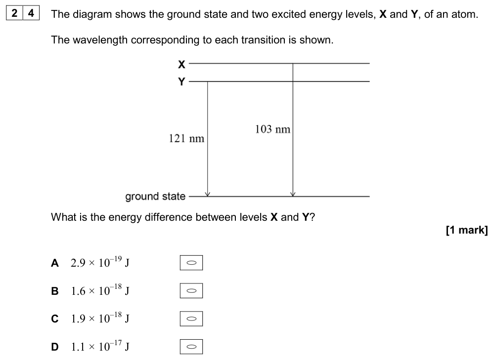
Photon energy for a transition
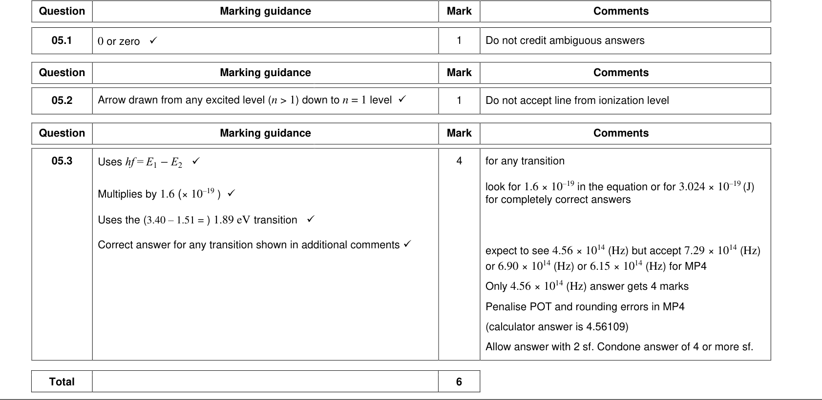
$$hf = E_1 - E_2$$
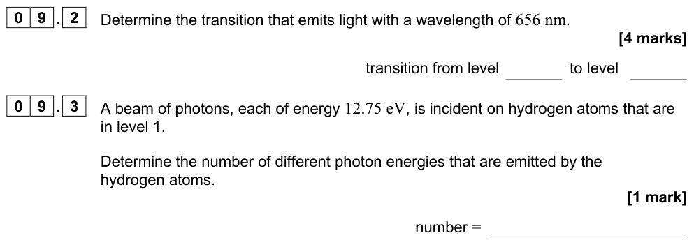
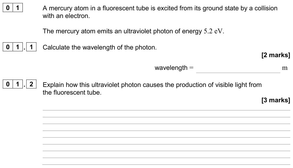
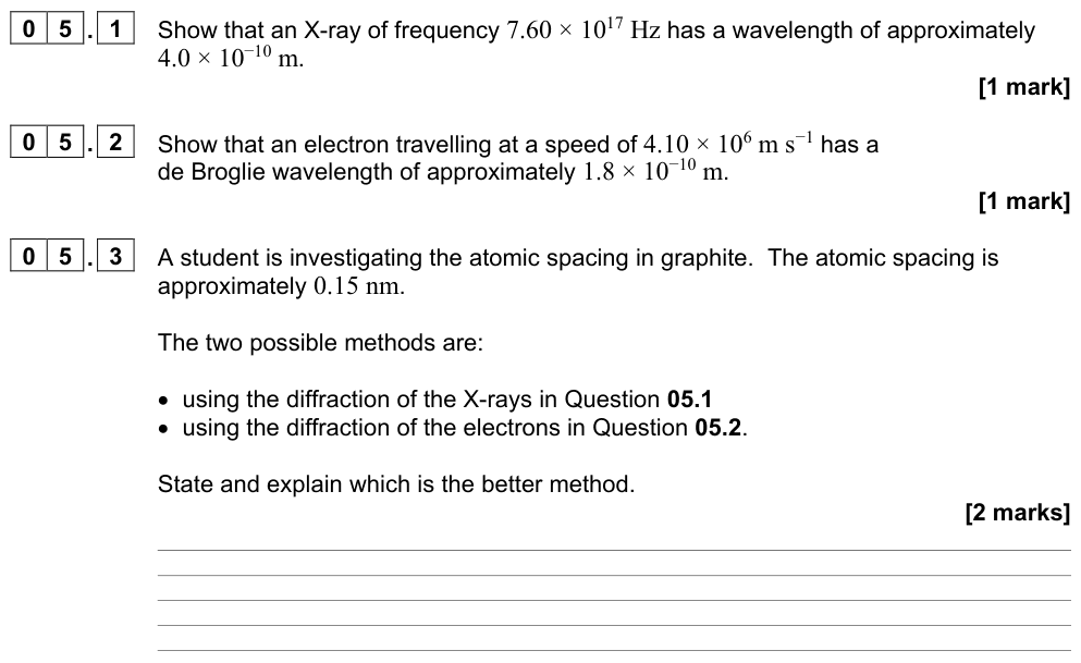
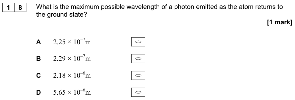
The photon energy equals the difference between the two energy levels. Since $E = hf = \dfrac{hc}{\lambda}$, a bigger energy gap gives a higher frequency and shorter wavelength.
The electronvolt
Electronvolt
$1\ \text{eV}$ is the energy transferred when an electron moves through a potential difference of 1 volt:
$$1\ \text{eV} = 1.60 \times 10^{-19}\ \text{J}$$
eV → J: multiply by $1.60 \times 10^{-19}$. J → eV: divide.
Worked example: hydrogen red line
An electron in hydrogen falls from $n = 3$ ($-1.51$ eV) to $n = 2$ ($-3.40$ eV).
$\Delta E = 1.89\ \text{eV} = 1.89 \times 1.60 \times 10^{-19} = 3.02 \times 10^{-19}\ \text{J}$.
$\lambda = \dfrac{hc}{\Delta E} = \dfrac{6.63 \times 10^{-34} \times 3.00 \times 10^{8}}{3.02 \times 10^{-19}} = 6.58 \times 10^{-7}\ \text{m} = 658\ \text{nm}$ (red).
Exam tip: counting spectral lines
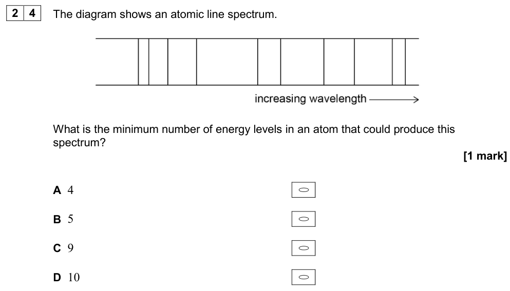
An atom excited to level $n$ can emit lines for every possible downward jump between levels. For $n$ levels the number of different lines is $\dfrac{n(n-1)}{2}$: from level 4, there are $4 \times 3/2 = 6$ lines.
Line spectra: evidence for energy levels
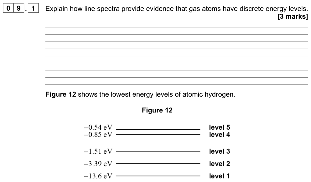
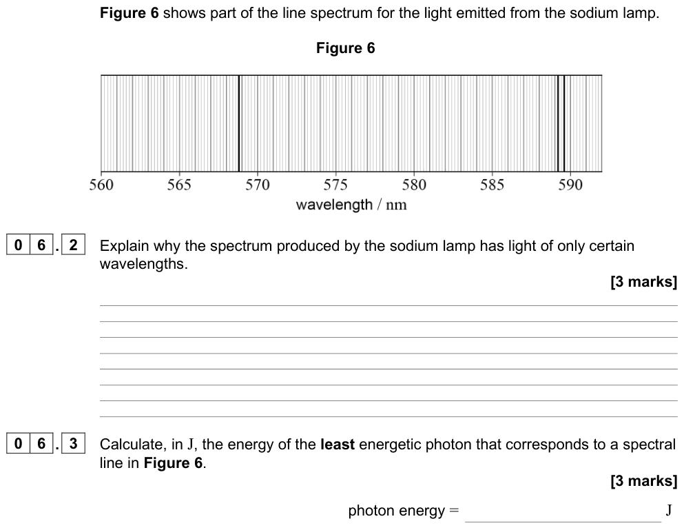
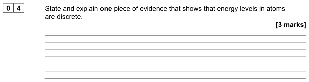
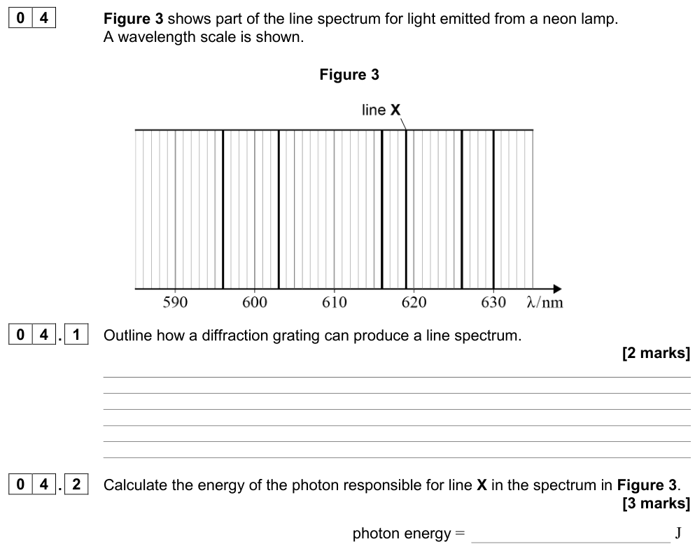
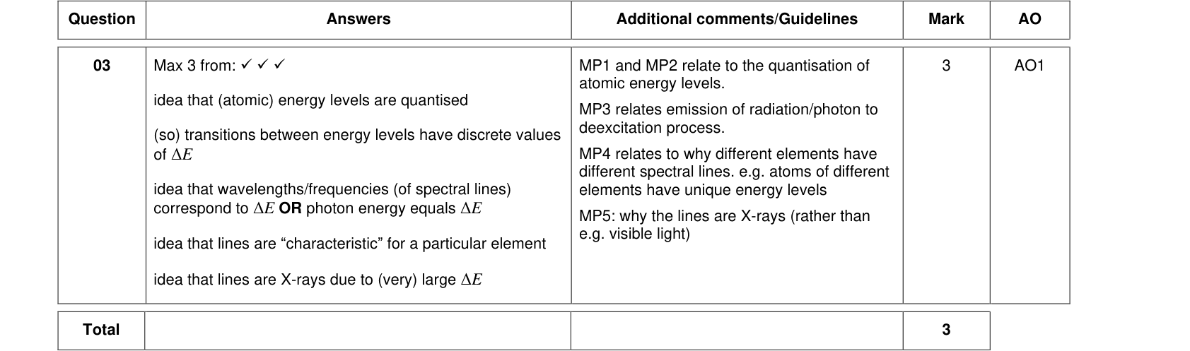
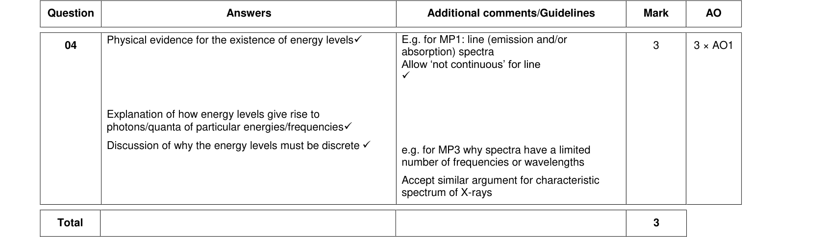
- An emission line spectrum (bright lines on a dark background) is produced by a hot, low-pressure gas. Each line corresponds to a particular transition between two energy levels and so has a particular, fixed wavelength.
- An absorption line spectrum (dark lines across a continuous spectrum) is produced when white light passes through a cool gas. The atoms absorb photons of exactly the energies needed for transitions, removing those wavelengths.
Because only certain specific wavelengths appear, the photon energies (and so the energy changes in the atom) are discrete. This is evidence that the electrons occupy discrete energy levels. If the electron energies could take any value, the spectrum would be continuous. Each element has its own unique set of lines, which is how the composition of stars is found.
The fluorescent tube
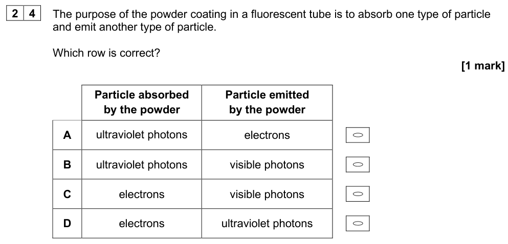
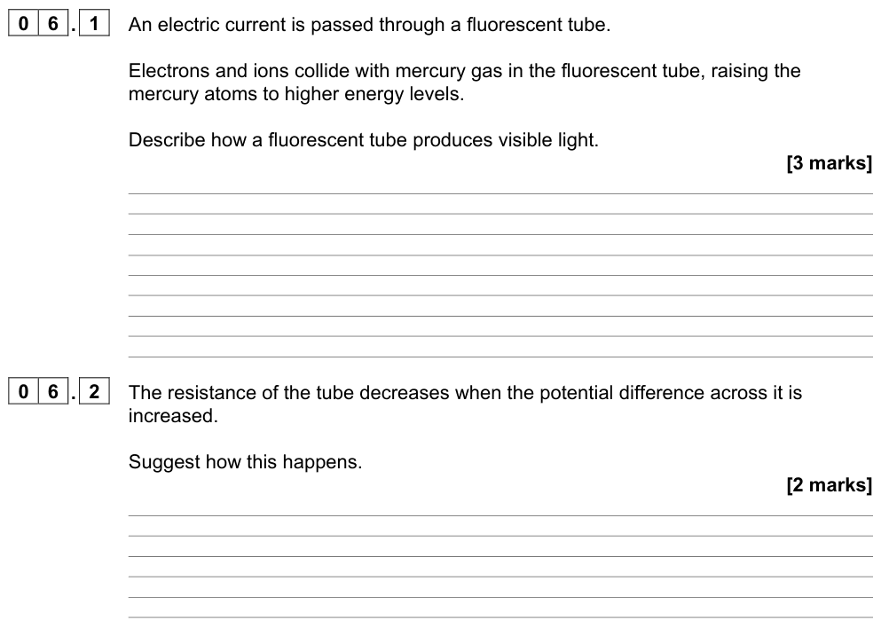
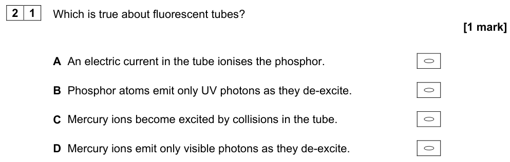
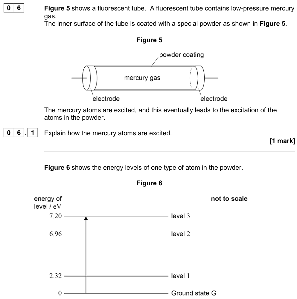
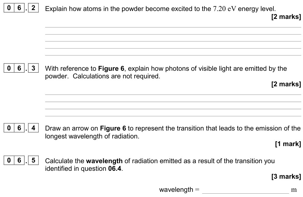
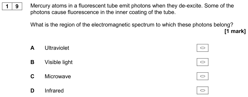
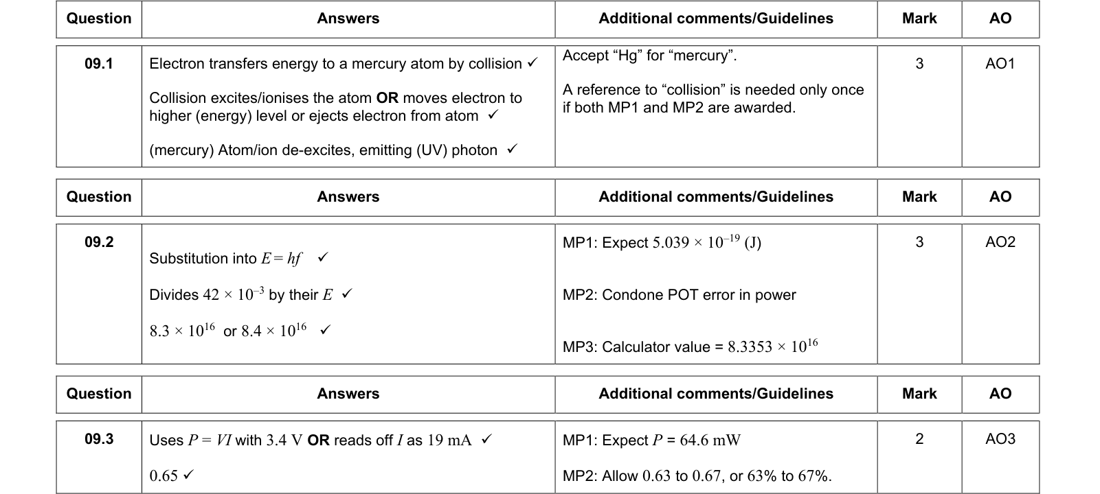
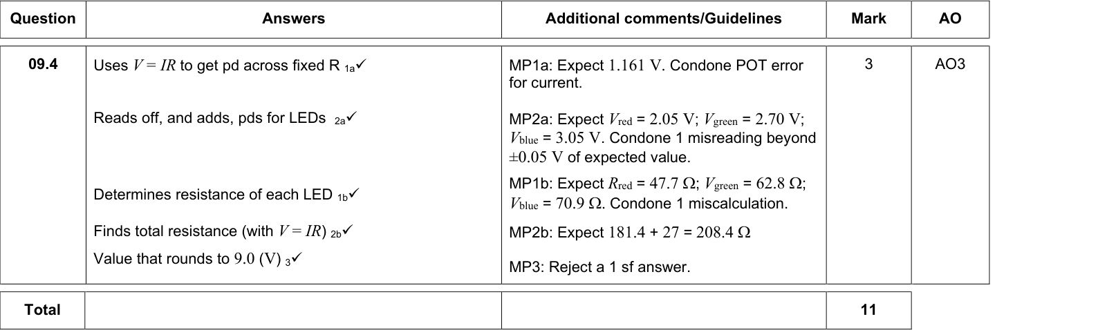
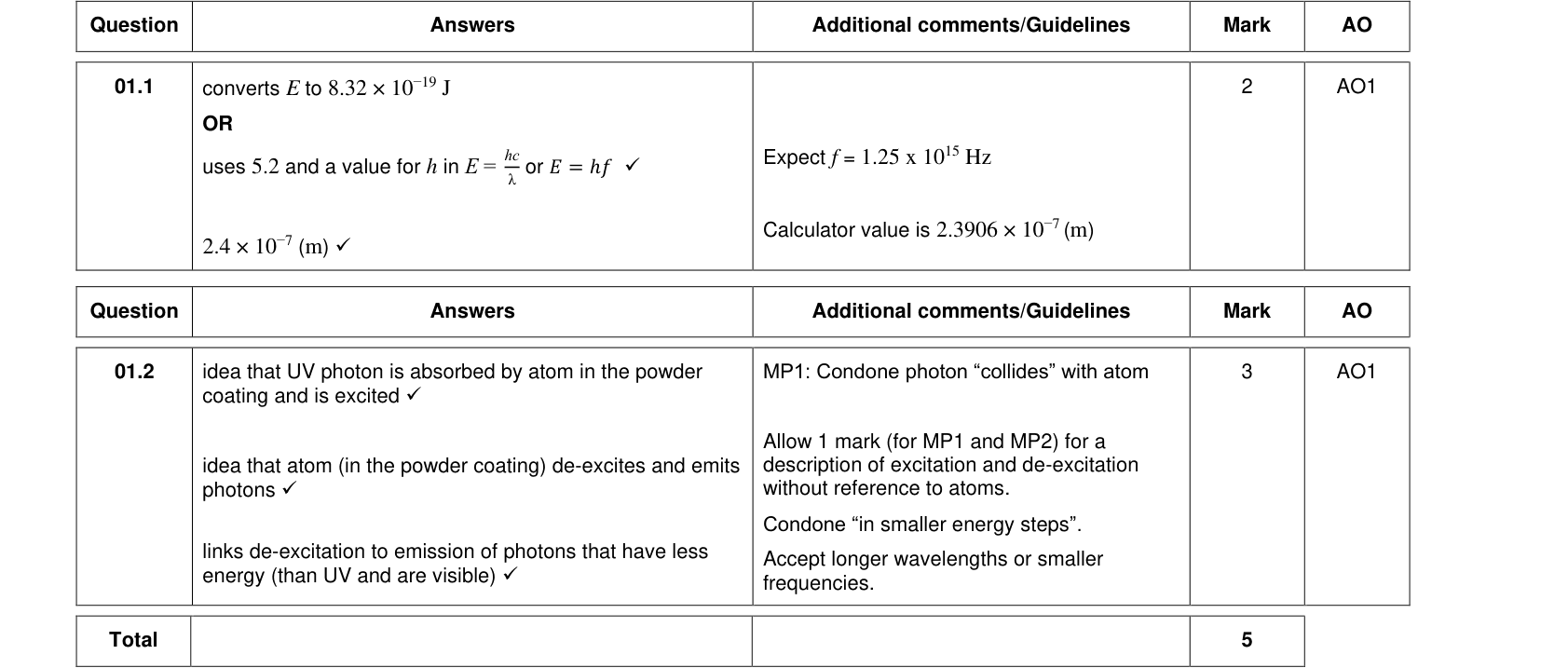
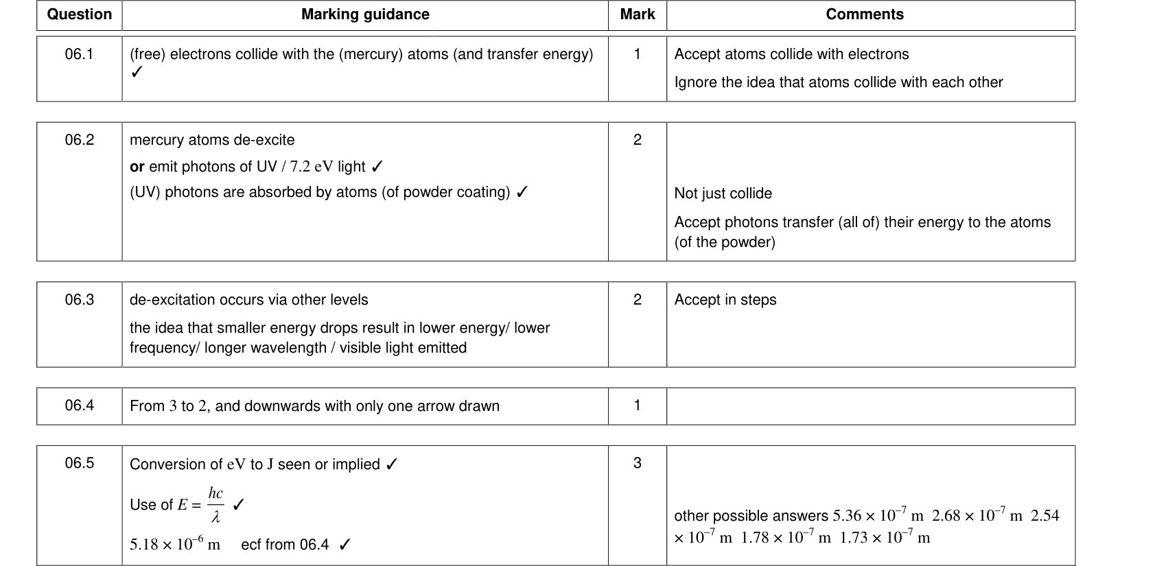
A fluorescent tube contains mercury vapour at low pressure. Its inside is coated with a phosphor (fluorescent powder).
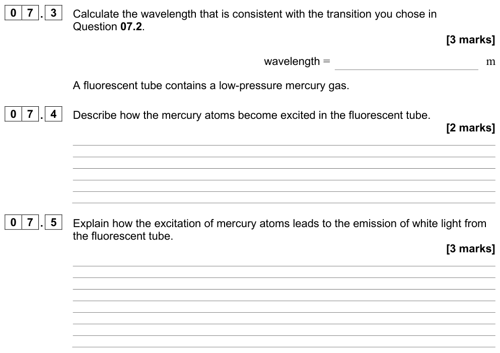
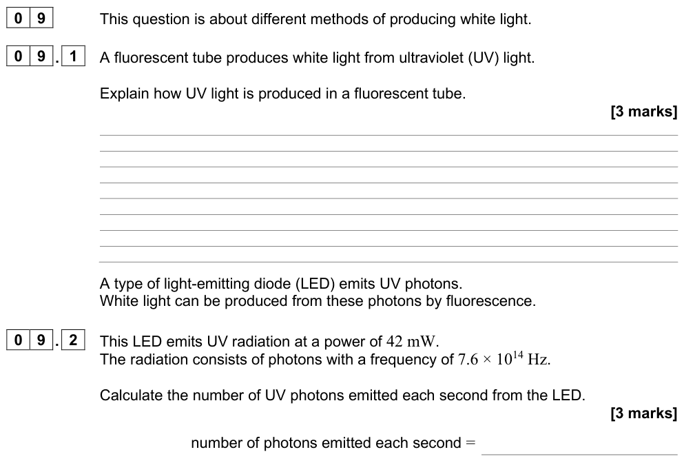
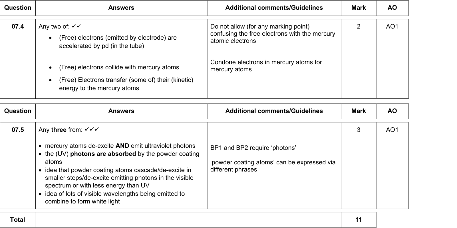
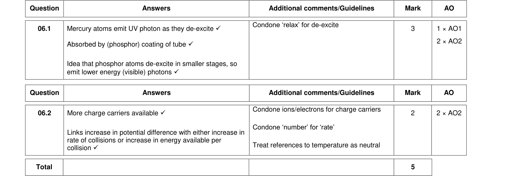
- A high p.d. across the tube accelerates free electrons from the cathode.
- The fast electrons collide with mercury atoms, exciting them (and ionising some, which keeps the current flowing).
- The mercury atoms de-excite, emitting photons, mostly in the ultraviolet (invisible and harmful).
- The UV photons are absorbed by atoms in the phosphor coating, exciting them to high levels.
- The phosphor atoms de-excite in several smaller steps, emitting lower-energy photons in the visible range.
The mix of phosphors is chosen so that the emitted light appears white.
X-rays
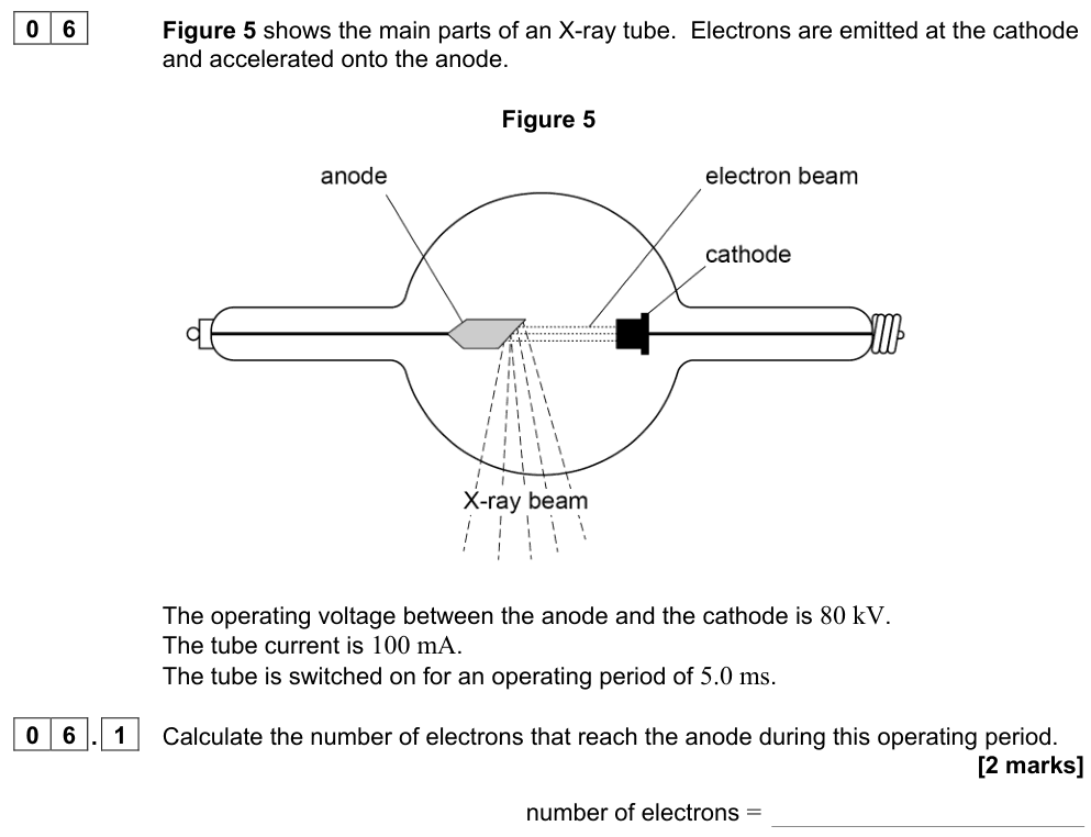
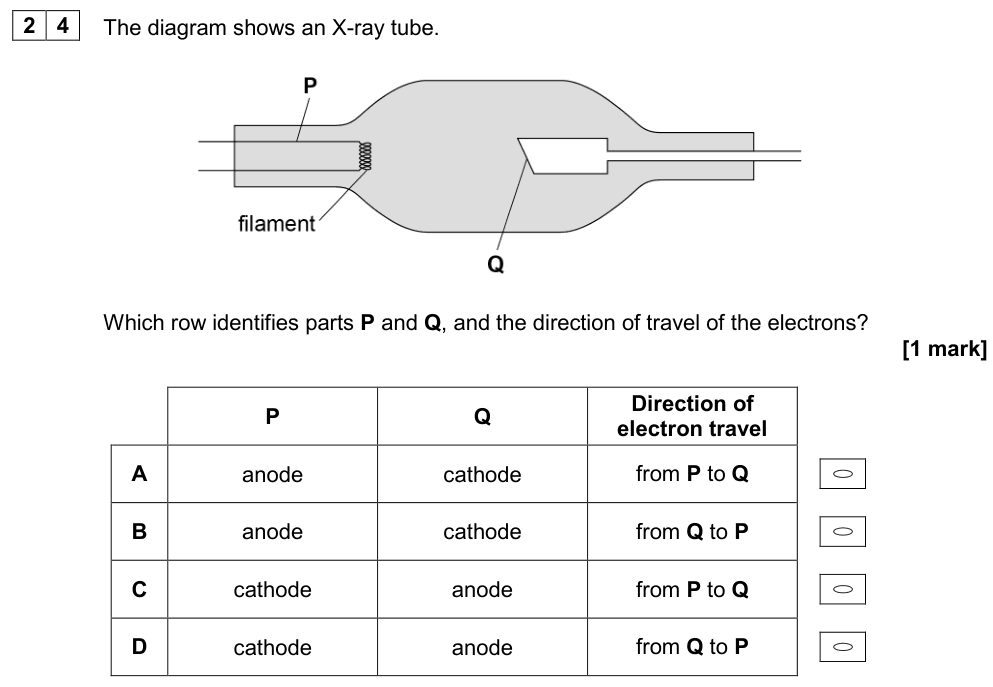
The X-ray tube
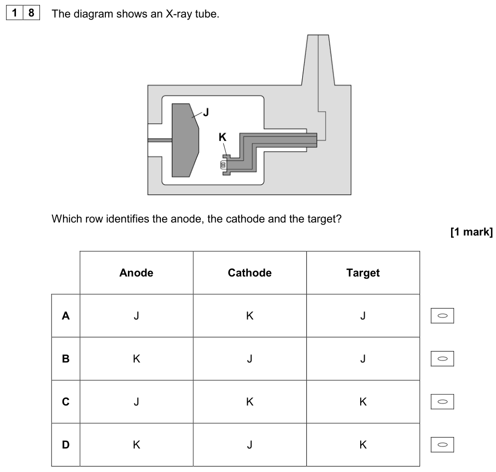
- A heated filament (cathode) releases electrons by thermionic emission.
- A very high p.d. (tens to hundreds of kV) between cathode and anode accelerates the electrons across an evacuated tube (so they do not collide with gas molecules).
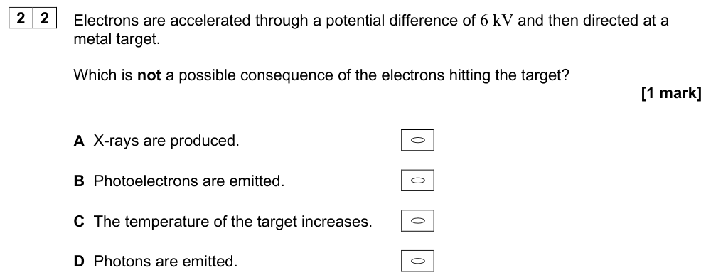
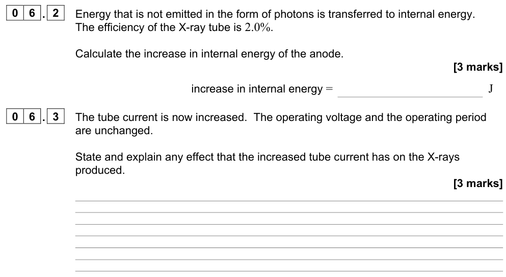
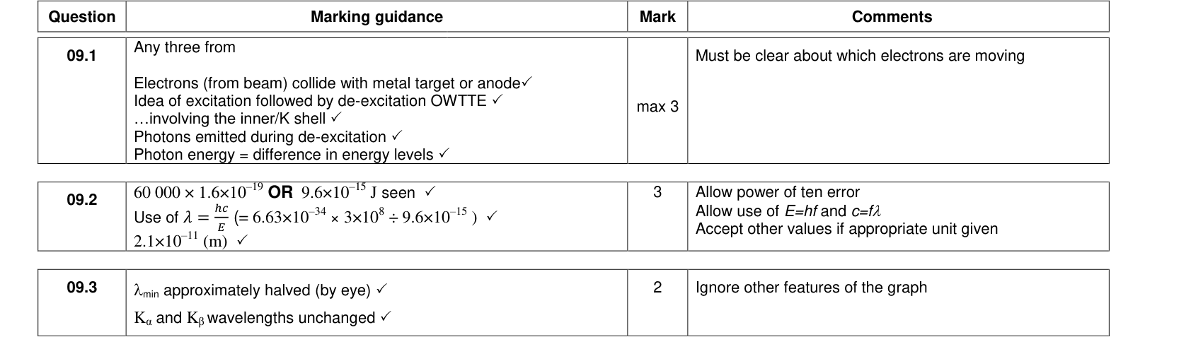
- The electrons strike a target of a heavy metal (usually tungsten, with a high melting point) on the anode. About 99% of their energy becomes thermal energy, so the anode is cooled (often by rotating it and by oil). About 1% becomes X-rays.
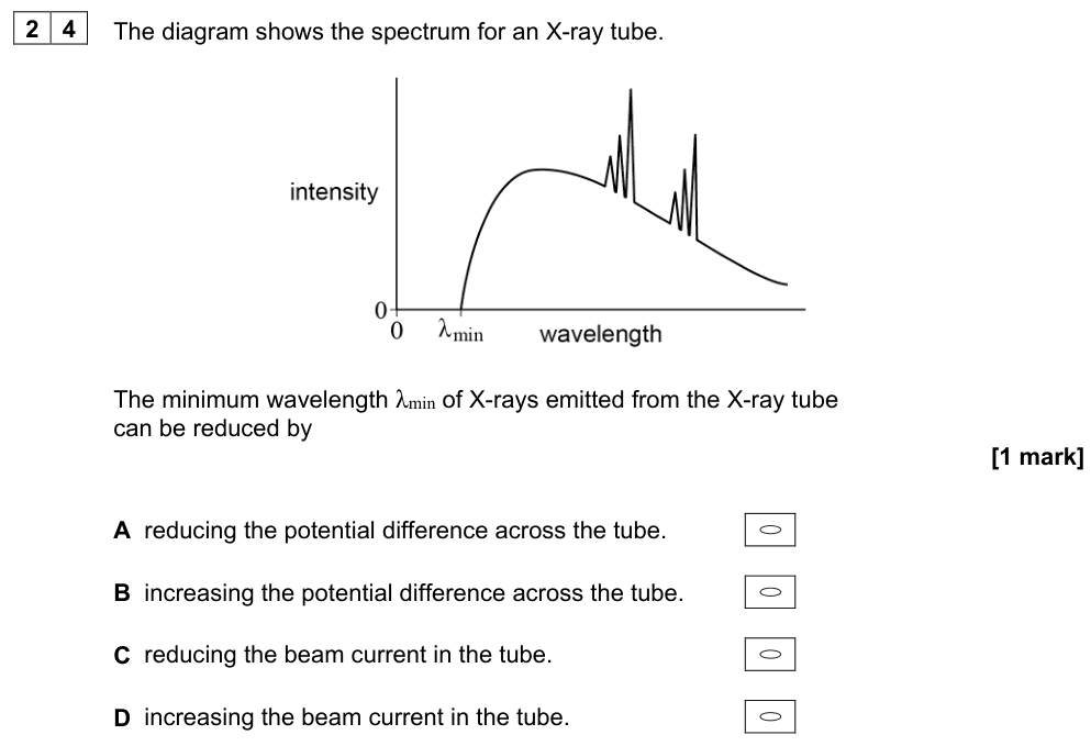
The X-ray spectrum
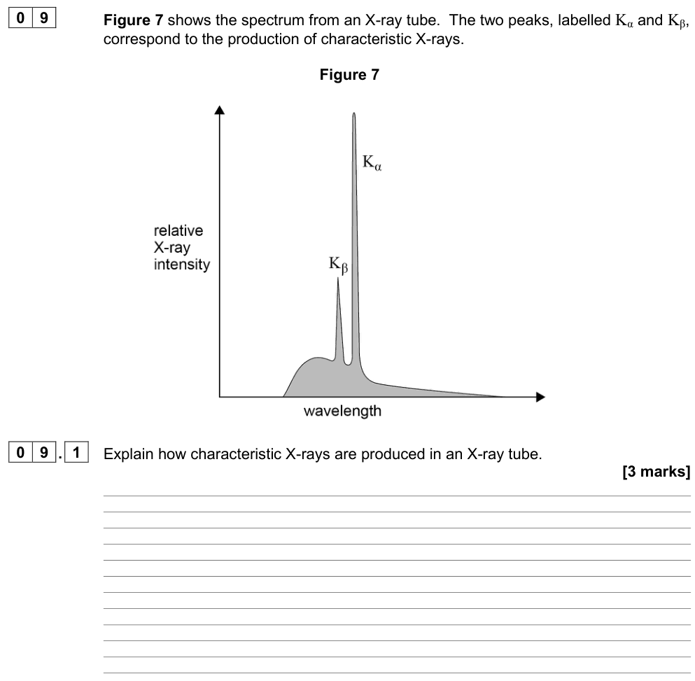
The spectrum has two parts:
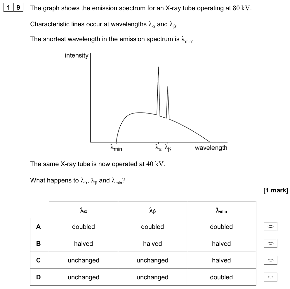
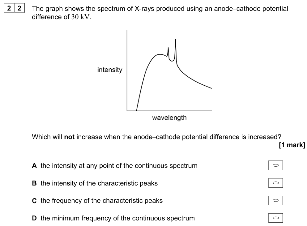
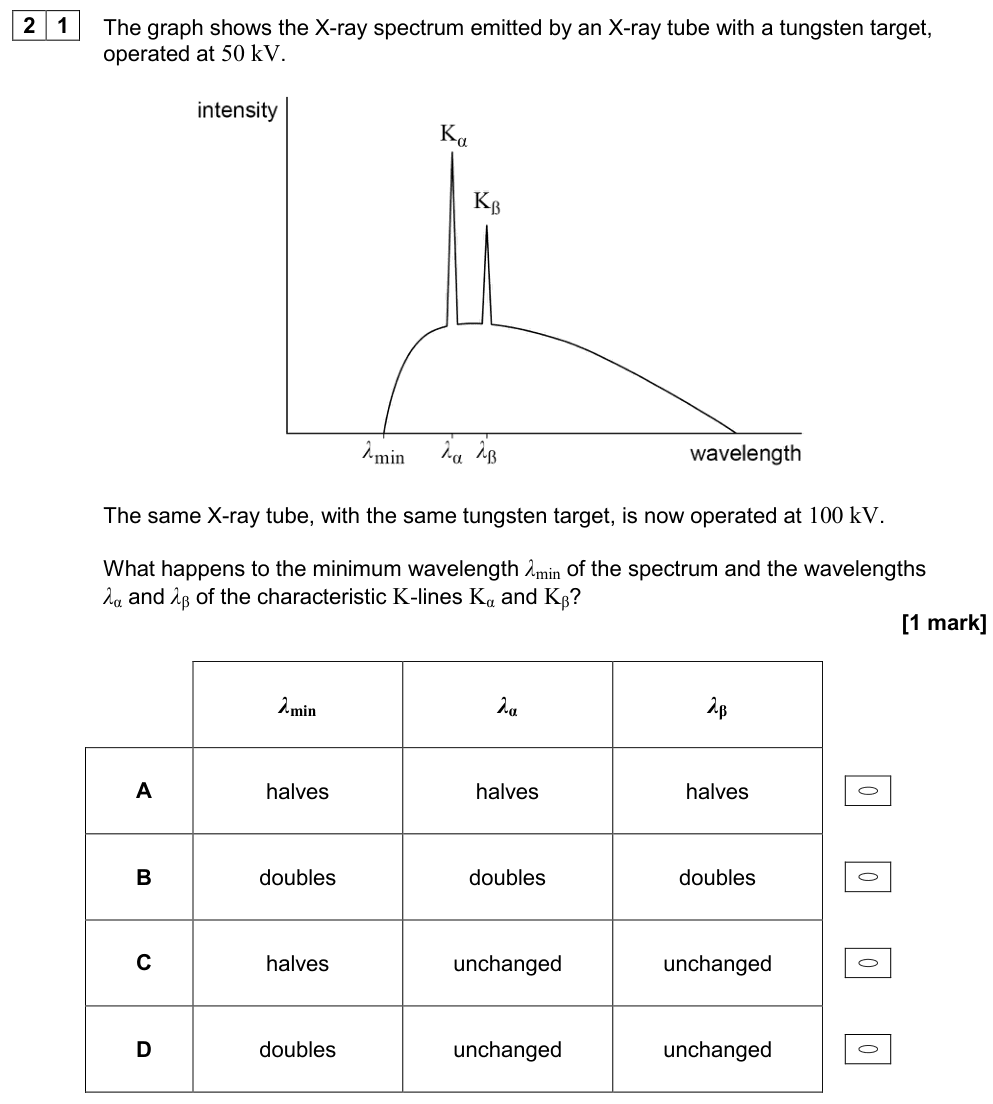
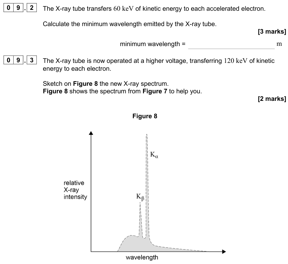
- A continuous spectrum (braking radiation, "bremsstrahlung"): electrons decelerate in the electric field of the target nuclei and lose kinetic energy as X-ray photons of any energy up to a maximum. The minimum wavelength corresponds to an electron losing all its kinetic energy $eV$ in one photon:
$$eV = \frac{hc}{\lambda_{\min}} \quad\Rightarrow\quad \lambda_{\min} = \frac{hc}{eV}$$
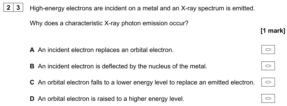
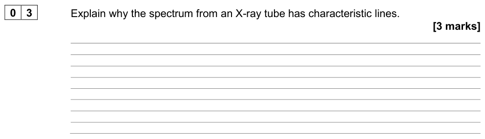
- Characteristic lines (sharp peaks): an incoming electron knocks out an electron from an inner shell of a target atom. An electron from a higher shell drops into the vacancy, emitting an X-ray photon with energy equal to the difference between the two inner-shell energy levels. The wavelengths of these lines are characteristic of the target element.
Changing the tube settings
| Change | Continuous spectrum | Characteristic lines | $\lambda_{\min}$ |
|---|---|---|---|
| Increase the accelerating p.d. | more intense, extends to higher energies | same wavelengths (become stronger) | decreases |
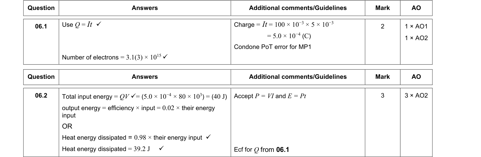
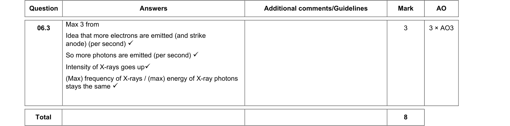
| Increase the filament (tube) current | more intense (more electrons) | same wavelengths, more intense | unchanged |
| Change the target metal | similar shape | different wavelengths | unchanged |
Medical uses of X-rays
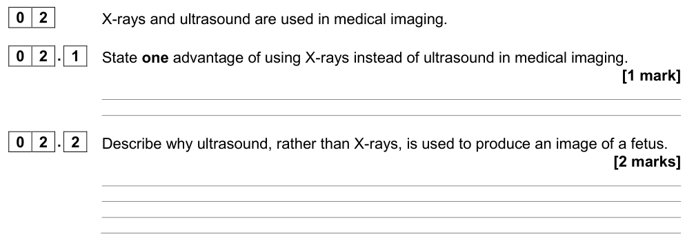
X-rays pass through soft tissue but are absorbed strongly by bone (dense, high proton number), so they produce shadow images of bones and teeth. CT scans combine many X-ray images from different angles to make cross-sectional images. High-energy X-rays are also used in radiotherapy to destroy cancer cells. X-rays are ionising, so the dose must be kept as low as possible; radiographers stand behind lead shields.
Summary
- Electrons occupy discrete energy levels; ground state lowest; ionisation level at 0 eV.
- Excitation (move up), ionisation (escape); photons are absorbed only if $hf$ = an energy gap; electrons can give part of their energy.
- Emission: $hf = E_1 - E_2$; line spectra prove that energy levels are discrete.
- Fluorescent tube: electrons excite Hg → UV → phosphor absorbs UV → re-emits visible light in smaller steps.
- X-ray tube: continuous spectrum with $\lambda_{\min} = hc/eV$ plus characteristic lines from inner-shell transitions.